What you need to know about cancer
Cancer is an uncontrolled growth of abnormal cells, which, if not diagnosed and treated in a proper way, can lead to tissue damage, metastasis, and ultimately death.
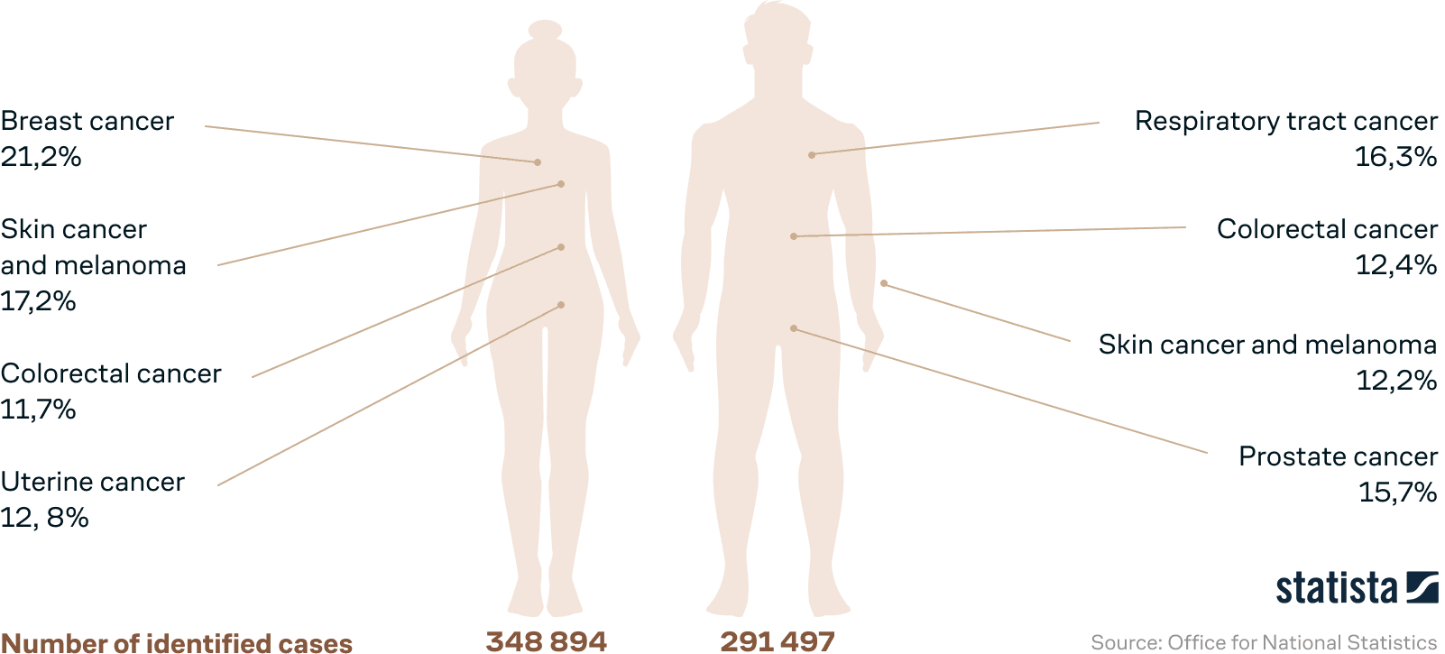
The most common cancers in Russia, 2019
There are over 100 types of cancer. In Russia the most common cancers of them are:
The most common types of cancer
In males
In females
In children
Symptoms
The typical symptoms of cancer will differ depending on the type and stage of the disease, but the most common include
- fatigue,
- weight loss,
- pain,
- skin changes,
- seals,
- disfunction of the internal organs,
- unusual bleeding,
- persistent cough,
- fever, etc.

Risk factors
Heredity
The presence of breast, ovarian, colon, prostate, skin cancer cases among close relatives, etc.
Infections
H. pylori, Epstein-Barr virus, HPV, hepatitis B and C viruses
Environmental factors
Air pollution, exposure to ionizing radiation and UV rays, exposure to chemical or toxic substances, etc.
Lifestyle
Smoking, drinking alcohol, malnutrition, obesity, physical inactivity, etc.
Elderly age
9 out of 10 cancers are diagnosed in people aged 45 and older
5 out of 10 cancers occur in people over 70
Causes
For cancerogenesis the cell must accumulate several breakdowns (mutations) in its DNA as a result of errors in division. Normally, the immune system recognizes and kills cells that differ from normal, but disturbances in the functioning of this system (stress, old age, immunodeficiency, etc.) can lead to the development of a cancer.


Formation of cancer


There are over 100 types of cancer
Lymphoma and myeloma
cancer that initiates in the the immune system cells. Multiple myeloma (MM) and lymphoma are both types that affect cells in a human blood. MM is a cancer of the plasma cells while lymphoma is a cancer of the lymphocytes. There are T-cell lymphomas, B-cell lymphomas, Hodgkin’s lymphomas, non-Hodgkin’s lymphomas, and lymphoproliferative lymphomas.
Leukemia (Leukemia)
cancer that starts in blood-forming tissue, such as the bone marrow, and causes large numbers of abnormal blood cells to form and circulate. There are lymphoblastic leukemias (ALL and CLL), myelogenous leukemias (AML and CML), T-cell leukemia and hairy cell leukemia.
Carcinoma
cancer that starts in the cells that line organs: the skin, lungs, colon, pancreas, ovaries. There are epithelial, squamous and basal cell carcinomas, melanomas, papillomas and adenomas.
Sarcoma
cancer that starts in bones, cartilage, adipose tissue, muscle, blood vessels, or other connective or supportive tissues — "bone, soft tissue cancer". Osteosarcoma, synovial sarcoma, liposarcoma, angiosarcoma, rhabdosarcoma and fibrosarcoma.
Сentral nervous system cancer
cancer that starts in tissues of the brain and spinal cord—gliomas, meningiomas, pituitary adenomas, vestibular schwannomas, primary CNS lymphomas, and neuroectodermal tumors.

The types listed above do not include metastatic tumor cells.
The reason is metastatic cells usually originate from the cell type named above, but end up in tissue from which they did not originally develop. Therefore, if the term "metastatic cancer" is used, then for greater accuracy it should include the tissue from which the tumor arises.
For example, a patient may say about having or being diagnosed with "metastatic cancer", but a more accurate statement is "metastatic (breast, lung, colon, or other type) cancer that has spread to the organ in which it was detected"
Another example: a doctor describing a man whose cancer has spread to the bones would say that he has metastatic prostate cancer in the bone.
This is not "bone cancer", which would be cancer that started in the bone cells. Metastatic prostate cancer in the bone is treated in a different way than lung cancer in the bone.


Hereditary forms of cancer
For example, the association between a pathogenic genetic variant and cancer development is well-studied for the following genes:

A Brief Comparison of Molecular Methods in Oncology
Molecular genetic methods are primarily used in oncology for diagnosis clarification, disease prognosis, and for selection of the most effective therapy (including targeted cancer therapy)
Next-generation sequencing (NGS)
is the most advanced and promising method that allows performing whole genome, whole exome, and targeted sequencing. Sequencing multiple DNA fragments occurs in parallel, providing high information content at a relatively high speed. NGS can simultaneously assess the presence of various genetic abnormalities in DNA and RNA molecules, such as hotspot mutations (mutations with increased frequency in the population), single nucleotide variants (SNV), copy number variations (CNV), small insertions, deletions, and gene fusions.
Oncofocus is an excellent example of an NGS assay used in cancer laboratory diagnostics.
«Oncofocus is a targeted next-generation sequencing (NGS), a multi-biomarker assay based on the Ion Torrent platform that detects variants across 52 cancer-relevant genes.
Pros
- A universal method that is able to detect a wide range of mutations
- Allows to perform diagnostics without prior hypothesis
- Multiple candidate genes can be analyzed simultaneously
- Only requires 10 ng of DNA or RNA
- One of the most sensitive methods
Cons
- High cost
- The method is used to diagnose only solid tumors
Other molecular genetic methods used in oncology
FISH — fluorescent in situ hybridization

FISH (fluorescent in situ hybridization) — allows you to determine the exact location of the nucleotide sequences on the DNA or RNA, visualizing them with complementary fluorescent DNA probes. FISH detects chromosomal rearrangements (deletions, duplications, translocations, inversions). In oncology, it is most applicable to clarify the diagnosis and to determine tumor status when selecting therapy.
Detects chromosomal aberrations: deletions, duplications, translocations, inversions
Pros
- Fast results
Cons
- Expedient to use when a mutation in a particular chromosome is suspected (cannot serve as a screening test)
- Requires confirmation of tumor molecular profile by other methods
- Unable to detect intra-chromosomal aberrations, i.e. most abberation types
- False positive results are possible (1-5%)
- High cost
- Detects chromosomal aberrations: deletions, duplications, translocations, inversions


arrayCGH — comparative genomic hybridization on microarrays
arrayCGH (comparative genomic hybridization on microarrays) is a method that detects unbalanced chromosomal rearrangements (deletions and duplications) of the entire genome. During the analysis, test and control DNA are labeled with different fluorescent dyes, mixed and hybridized on microarrays with DNA probes, quantitatively comparing the intensity of fluorescence.
Pros
- Allows to analyze the presence of pathogenic variants in the whole genome simultaneously
Cons
- Indicates only the presence of a variant and requires additional methods for clarification
- Unable to detect balanced chromosomal rearrangements and point nucleotide variants (replacements, deletions, insertions)
- Demanding to the incoming DNA concentration
- High cost
PCR — Polymerase Chain Reaction
PCR (polymerase chain reaction) is a simple and effective laboratory diagnostic method that allows multiple copies of specific DNA or RNA fragments to be multiplied for qualitative or quantitative assessment of the fragment sought. In oncology, it can be used to detect single-nucleotide genetic variants (allele-specific PCR) or microRNA. More often, however, PCR is part of more sophisticated diagnostic methods, such as sequencing. In addition, PCR is widely used to detect the presence of potentially carcinogenic pathogens (HPV types 16 and 18, EBV infections, HBV and HCV, retroviruses, etc.).
Detects the presence of potentially carcinogenic pathogens HPV types 16 and 18, EBV infection, HBV and HCV, retroviruses, etc.

Pros
- Relatively inexpensive
- Accessible
- Enables detection of potentially carcinogenic viral and bacterial DNA/RNA agents
- Allows use of any biomaterial containing DNA or RNA
Cons
- False positive results may occur
- In diagnostic laboratories the analysis is usually limited to searching for the most common mutations in a given population
- For best performance, it must be combined with other methods
Treatment
In cases where a malignant tumor is not operable, the patient receives combined treatment:
Combination of
- surgery,
- chemotherapy,
- radiotherapy,
- immunotherapy or targeted therapy.

Meanwhile, immunotherapy and targeted drugs are the most effective and, at the same time, safe method for healthy tissues.
Their use is based on the search for molecular biological markers of diagnosed cancer for the subsequent preparation of an effective treatment plan.